Three years after a group of Caltech researchers demonstrated that pairs of entangled photons could quadruple a light microscope’s resolution, the same lab has discovered a method to increase this improvement even more. By employing a novel optical design that passes one of the entangled photons via the microscope’s optics three times instead of just once, they have now attained a fourfold increase in resolution when compared to a traditional microscope. In a study published in the journal Science Advances, the team details the setup and experimental validation.
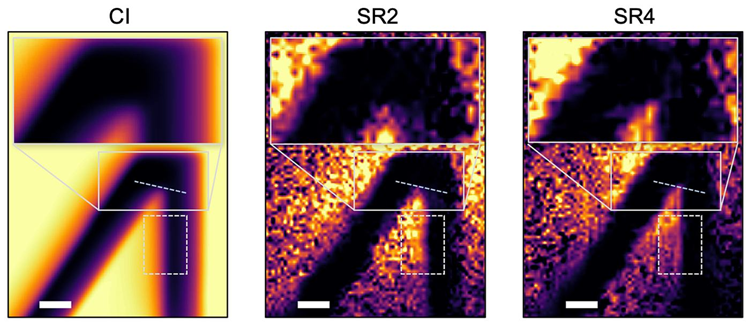 Comparing images of a resolution target using classical microscopy (left), twofold super-resolution imaging (SR2) where the idler beam only passes once, and fourfold super-resolution imaging (SR4) where the idler beam passes back and forth three times. Notice how much more detail in the image is provided by the SR4 quantum imaging technique. The scale bars represent 10 µm. Image Credit: California Institute of Technology
The research, directed by Lihong Wang, the Bren Professor of Medical Engineering and Electrical Engineering and the Andrew and Peggy Cherng Medical Engineering Leadership Chair at Caltech, advances on the lab’s 2023 demonstration of quantum microscopy by coincidence (QMC). The technique is based on entanglement, a strange quantum-mechanical phenomenon in which two particles are connected so that the state of one particle is inextricably linked to the state of the other, regardless of how far away they are.
In QMC, entangled pairs of photons, known as biphotons, are separated so that one photon, the signal photon, goes through the sample while its entangled counterpart, the idler photon, travels a distinct parallel route. In certain aspects, the pair behaves like a single particle, with twice the momentum of a single photon.
According to quantum mechanics, a particle's wavelength is inversely related to its momentum. This means that one of the photons in a biphoton pair can be successfully imaged with a wavelength half that of the original light. And because microscope resolution improves as wavelength reduces, this results in a twofold increase in resolution.
In the new system, the team transmits the signal photon through the object being imaged only once. The idler photon, however, passes through the same pair of lenses three times before reaching the detector. To achieve this, the researchers generate a magnetic field and use optical components, including specialized beam splitters, to manipulate a property of light known as polarization, essentially the direction in which a light wave’s electric field oscillates.
Now we have entered a new physical regime. In general, people think that with a single photon pair, you can, at most, increase the resolution from the classical diffraction limit by two times. But we've gone beyond that. This is the most exciting aspect of our new paper, because it points us in a direction where we can improve even further – 10 times or maybe even 100 times – down the road.
Lihong Wang, Bren Professor, Medical Engineering and Electrical Engineering, California Institute of Technology
Wang and his colleagues tested the new system’s capabilities by imaging a standard test target using a classical imaging setup, the previous twofold quantum configuration, and the new triple-pass configuration. By comparing the sharpness of the target’s edges and fine details, they found that the twofold setup improved resolution by about 1.8 times compared with classical imaging, while the new triple-pass system delivered an improvement of nearly four times.
The study also has significant biomedical implications. Conventional high-resolution retinal imaging can require light intense enough to temporarily impair a patient’s vision. In the new quantum imaging approach, however, the light that reaches the imaging target is far less intense, reducing the risk of damage to living tissue. According to the researchers, higher-resolution imaging at low light levels could eventually make it possible to observe much smaller structures, such as cell nuclei and mitochondria, without causing damage.
It might also be beneficial in semiconductor device inspection, where quality-control imaging sometimes requires longer wavelengths, which result in low resolution with classical optics. According to Wang, a fourfold improvement in resolution at the same wavelengths might significantly improve defect identification in chips.
Wang emphasizes that, while the experimental results verify the novel strategy, his team is still working on a theoretical model to explain the technique's potential to improve resolution. Wang admits that initially, he struggled to persuade his lab to pursue this approach.
Based on our previous theory, I thought this might be possible, but it was just a hunch. People felt it was risky, but eventually they started working on it. One night, one of my postdocs emailed me and said, 'It worked!' It was such a great moment. And now that we've observed this, we definitely want to push it even further.
Lihong Wang, Bren Professor, Medical Engineering and Electrical Engineering, California Institute of Technology
The study, titled “Above twofold quantum super-resolution microscopy enabled by multiple idler passes with entangled biphotons,” was written by Wang lab alumnus Xin Tong (PhD '26), former postdocs Zhe He (now at Shandong Institute of Advanced Technology in China), and Yide Zhang (now at the University of Colorado Boulder).
The study's authors include Wenyu Liu and Chien-Ying Huang (MS '23), graduate students at Caltech. The National Institutes of Health, the Silicon Valley Community Foundation, the Chan Zuckerberg Initiative DAF, and Caltech's Center for Sensing to Intelligence provided funding for the study.
Journal Reference:
Tong, X., et al. (2026) RAbove-twofold quantum super-resolution microscopy enabled by multiple idler passes with entangled biphotons. Science Advances. DOI:10.1126/sciadv.aea9457. https://www.science.org/doi/10.1126/sciadv.aea9457.