Image Credit: Micha Weber/Shutterstock
LCM, put simply, is a form of microscopy which utilizes laser excitation, fluorescent light and optical screening techniques to produce a highly refined, digital image. This form of microscope was first theorized and later developed by Marvin Minsky, a co-founder of the Massachusetts Institute of Technology’s artificial intelligence department and a leading researcher of his day.
Minsky developed the first confocal microscope in 1955 and later patented the idea in 1957. Unfortunately, the full potential of his invention would not be realized for several decades, as technology caught up and digital imaging came into use.
Why is Confocal Microscopy Notable?
LCMs can produce renderings of samples which in practice break what would normally be considered the resolution limit of a non-electron-based microscope. This is due to their ability to selectively detect light on specific focal planes, effectively removing background, unfocussed, light from the image. This is achieved through optical screening, by using a minute pinhole-size aperture, linked to a photomultiplier tube and digital display.
How Does Laser Confocal Microscopy Work?
First, a sample will undergo fluorescent tagging, utilizing a fluorescent protein marker which will be genetically spliced into the DNA of the sample. Notably, multiple fluorescent tags can be used respectively for different molecules, which is key in differentiating between structures within the sample under study.
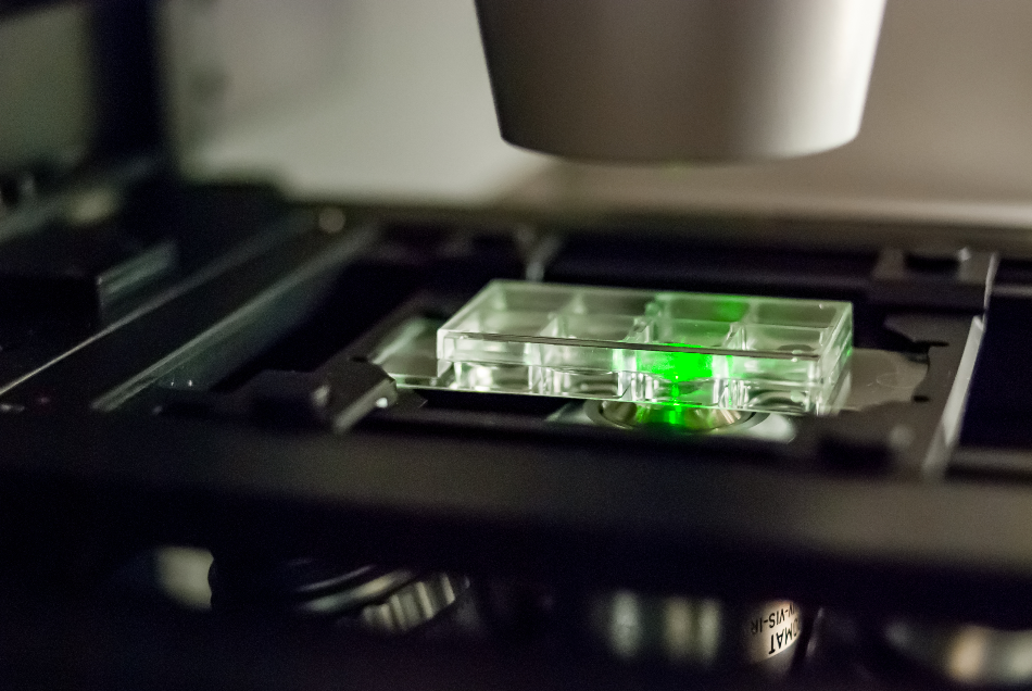
A section of the sample will then be prepared and placed on the observation platform of the microscope. From here, a highly focussed laser beam will then be directed at the sample, via reflection and refraction from a series of mirrors. This then causes the tags to fluoresce, emitting a wavelength of light discrete from the other tags and the excitatory beam.
This fluorescent light is then relayed back to a computer. However, this journey is more complex than it might at first seem. When the laser beam is targeted at the sample, multiple molecules will fluoresce, creating a ‘mess’ of emitted light.
The entirety of the fluorescent light, from all tags in the sample, is reflected off a dichromatic mirror which directs it effectively towards an optical screen with a pinhole aperture. This tiny aperture allows light from a targeted focal plane to be isolated from the rest of the light which has been produced from the sample. The now focussed beam of light is then targeted to a photomultiplier tube, which perceives the photons via a special photocathode.
The minute and isolated fluorescent signal is magnified inside the photomultiplier, being passed along the tube from dynode to dynode and then to a terminal anode. An output meter then detects the signal.
From here, the signal is perceived as an electrical signal by a PC program and can be reassembled as a single-pixel within a picture.
What Does This Mean?
Due to the way in which an LCM renders an image, it is a slower form of microscopy than the traditional widefield or brightfield microscopy. This is because at any one time only a single microscopic point of a sample is being visualized and a full rendering must, therefore, be constructed pixel by pixel. The result is a slow process and incredibly detailed visualization of a specific target structure within the object under study.
Why is LCM an Advantageous Form of Microscopy?
Alongside the remarkable visualization and resolution potential of LCM, comes the amazing benefit of 3-D image visualization. Unlike a scanning electron microscope which effectively gives a 3D visualization of the surface ‘landscape’ of a target sample, an LCM is capable of sequentially scanning thin layers of the sample to produce a 3-D model of an internal structure, such as an organelle. When this is combined with multiple fluorescent tags the resultant image can be built to incorporate many structures to show their interplay and individual structures.
As an added advantage, utilizing different colored fluorescent tags allows an LCM to produce a colored image to aid in structural differentiation, without the need for the application of false color later. As such, LCM technology has been revolutionary in the fields of microscopy and microbiology and is now a staple piece of kit in any state-of-the-art research facility.
Sources and Further Reading
Disclaimer: The views expressed here are those of the author expressed in their private capacity and do not necessarily represent the views of AZoM.com Limited T/A AZoNetwork the owner and operator of this website. This disclaimer forms part of the Terms and conditions of use of this website.