Photon-Counting Computed Tomography (CT) is an innovative new technology that has remarkable potential to change clinical CT.

Image Credit: sKjust/Shutterstock.com
CT is a diagnostic imaging method used to produce high-resolution images of internal organs, bones, soft tissue, and blood vessels. Sir Godfrey Hounsfield invented the first CT scanner using x-ray technology in 1967. The first clinical use of a CT scanner is historically identified to have been performed in 1971. Since then, CT technology has advanced through many improvements.
Initially, single-section CT scanners were introduced. Later, multidetector systems capable of imaging entire human organs were developed. Further evolutions reduced the imaging time and the size of CT scanners. Highly efficient algorithms and reconstructive models have been developed to complement the growing technology.
Limitations in Conventional CT Technology
Although CT technology has been immensely successful in advancing clinical medicine, it still has considerable limitations. For example, the ionizing radiation produced by the x-ray beams in a CT scanner can have harmful effects on patients. If the amount of exposed radiation is reduced, the image quality suffers from distortions. Poorer detection quality leads to insufficient details of the sample studied.
Another drawback of conventional CT images is the low contrast between different types of soft tissues. This limitation fails to reliably differentiate between pathologic and healthy tissues.
A technique that provides better images that can identify specific tissues as well as mitigate some other health risks associated with conventional CT methods is photon-counting CT.
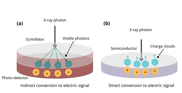
Figure 1: Schematic illustrations showing the operating principle of (a) conventional CTs using EIDs and (b) photon-counting CTs. Image Credit: Ilamaran Sivarajah
Photon-Counting CT
Photon-counting CT uses different types of detectors to resolve x-ray light, a mechanism that is fundamentally different from the type of detectors used in conventional clinical CT.
Conventional CT scanners use energy-integrating detectors (EIDs). An EID consists of a detector bank. A typical EID detector bank consists of an array of detector elements. The total x-ray energy deposited on each detector element is measured for a given time. As seen in figure 1 (a), in conventional CTs, incident x-rays are absorbed on the top layer.
The top layer, called the scintillator, converts x-ray photons into visible light. A shower of secondary visible light photons is generated when x-ray photons strike the scintillator. The bottom layer of the detector is a photodiode made of semiconducting material. The spray of visible photons is converted to an electrical signal by the photo-detector. The electrical signal is proportional to the total energy deposited during the measurement interval. The energy of an individual x-ray photon cannot be measured using this method.
More from AZoQuantum - Massive Black Holes: Where Do They Come From?
Photon-counting CT technology is equipped with a photon-counting detector (PCD). PCDs underlying operational mechanism is different from EIDs. PCDs do not rely on the scintillator layer to convert x-ray photons to visible photons. Instead, PCDs are constructed with one layer of a semiconductor diode. A large voltage is applied to create an electrical bias between the top and bottom of the photo-diode. When an incident x-ray photon is absorbed by the semiconductor, it generates a cloud of positive and negative charges.
The charges rapidly pull away from each other due to their repulsive force. Wires that are attached to the photo-diode transmit the electrical pulse generated by the moving charges. The electrical pulse is recorded with an electronic circuit. The electrical signal generated by an individual detector element in a PCD is illustrated in figure 1 (b). PCDs convert individual x-ray photons directly into an electric signal.
This is an advantage over conventional CTs that use EIDs which require the additional step of converting photons to visible light. Apart from the direct conversion of x-ray photons, PCD’s also provide additional benefits. These include no electric noise, smaller detector pixels, and intrinsic spectral imaging built into each scan.
Challenges
There are several technological challenges that have to be overcome for optimal PCD performance. For example, cross-talk has to be reduced. Cross-talk happens in a PCD when more than one detector element registers a signal for the same incoming x-ray photon.
Another drawback of PCD is pile-up. Pile-up occurs when the detector is not fast enough to individually resolve all the x-ray photons impinging on the detector. Several hundred million photons impinge on the detector per second per square millimeter. Failure to rapidly measure each photon results in pile-up.
Images Taken with Photon-Counting CTs
Various academic research organizations have developed and implemented photon-counting CTs. Such images were produced in the research group of Dr. Anders Persson at Linkoping University in Sweden. The middle and inner ear were imaged using conventional CT and photon-counting CT.
Small anatomical structures of the middle and inner ear such as the stapes and the cochlea were captured. Images taken with conventional CTs and photon-counting CTs were taken and compared, with photon-counting CTs clearly showing much higher resolution.
Future Outlook
Photon-counting CT detectors were approved for clinical use by The U.S. Food and Drug Administration (FDA) in September 2021. With the capabilities provided by this technique, like reduced radiation exposure and higher resolution images, photon-counting CT is a powerful medical imaging tool. It promises to improve diagnosing diseases, trauma, as well as planning, guiding, and monitoring interventional or therapeutic procedures.
References and Further Reading
Martin J. Willemink, Mats Persson, Amir Pourmorteza, Norbert J. Pelc, and Dominik Fleischmann, “Photon-counting CT: Technical Principles and Clinical Prospects.” Radiology 2018 289:2, 293-312 https://pubs.rsna.org/doi/10.1148/radiol.2018172656
VIDEO: Clinical CT Applications with Photon Counting Detectors — Reuven Levinson, GE Healthcare, Haifa, ISRAEL.
First Photon-counting CT System Cleared by the FDA. (2021, November 9). DAIC. https://www.dicardiology.com/article/first-photon-counting-ct-system-cleared-fda
Disclaimer: The views expressed here are those of the author expressed in their private capacity and do not necessarily represent the views of AZoM.com Limited T/A AZoNetwork the owner and operator of this website. This disclaimer forms part of the Terms and conditions of use of this website.